New case emerging for Culex mosquito as unexpected Zika spreader
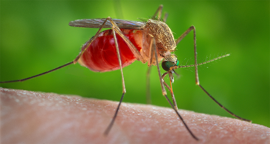
ORLANDO, Fla. — New evidence from separate labs supports the controversial idea that an overlooked and unexpected Culex mosquito might spread Zika virus.

The southern house mosquito, Culex quinquefasciatus, is common in the Americas. Constância Ayres, working with Brazil’s Oswaldo Cruz Foundation in Recife, previously surprised Zika researchers with the disturbing proposal that this mosquito might be a stealth spreader of Zika. But two U.S. research groups tested the basic idea and couldn’t get the virus to infect the species.
Now, preliminary results from Ayres’ and two other research groups are renewing the discussion. The data, shared September 26 at the International Congress of Entomology, suggest that Zika can build up in the house mosquito’s salivary glands — a key step in being able to transmit disease. Basic insect physiology is only part of the puzzle, though. Even if the mosquitoes prove competent at passing along Zika, there remain questions of whether their tastes, behavior and ecology will lead them to actually do so.

In the current outbreak, the World Health Organization has focused on mosquitoes in a different genus, Aedes, particularly Ae. aegypti, as the main disease vector. But Ayres had announced months ago the discovery of the virus in Brazil’s free-flying house mosquitoes (SN Online: 7/28/16).

At the congress, Ayres’ foundation colleague Duschinka Guedes reported that captive mosquitoes fed Zika-tainted blood had virus growing in their own guts and salivary glands within days. The virus doesn’t spread every time a mosquito slurping contaminated blood gets virus smeared on its mouthparts, though. To move from the mosquito to what it bites, viruses have to infect the insect midgut, then travel to the salivary glands and build up enough of a population for an infective dose drooling into the next victim. When Guedes offered the infected mosquitoes a special card to bite, they left telltale virus in the salivary traces, a sign of what they could do when biting — and infecting — a real animal.

Researchers from China and Canada who were not originally on the symposium program also stepped up to share their results, some of which are unpublished. Some tasks are still in early stages, but both labs showed Zika virus building up in some kind of Culex mosquitoes.

At the Beijing Institute of Microbiology and Epidemiology, Tong-Yan Zhao found the virus peaking in the house mosquitoes eight days after their first contaminated drink. As a test of the infectious powers of the mosquitoes, researchers let the Zika-carrying insects bite baby lab mice. Later, the virus showed up in the brains of eight out of nine lab mice. The results were reported September 7 in Emerging Microbes & Infections.
From Brock University in St. Catharines, Canada, Fiona Hunter has found signs that 11 out of 50 wild-caught Culex pipiens pipiens mosquitoes picked up the virus somewhere on their bodies. So far, she has completely analyzed one mosquito and reports that the virus was indeed in its saliva.

These positive results contradict Culex tests at the University of Texas Medical Branch in Galveston. Those tests, with U.S. mosquitoes, found no evidence that C. quinquefasciatus can pick up and pass along a Zika infection, says study coauthor Scott Weaver. Stephen Higgs of Kansas State University in Manhattan and his colleagues got similar results. “We’re pretty good at infecting mosquitoes,” Higgs says, so he muses over whether certain virus strains won’t infect mosquitoes from particular places.

The main risk from Culex at the moment is distraction, warned Roger Nasci of North Shore Mosquito Abatement District in Northfield, Ill. After the talks, he rose from the audience to say that Ae. aegypti is a known enemy and limited resources should not be diverted from fighting it.

George Peck, who runs mosquito control for Clackamas County in Oregon, isn’t convinced that the high virus concentrations dosing the test mosquitoes are realistic. Yet he’s watching the issue because like much of northern North America, Clackamas doesn’t have the Ae. aegypti vector to worry about. But it does have plenty of Culex mosquitoes.

Nerve cell migration after birth may explain infant brain’s flexibility

Baby humans’ brain cells take awhile to get situated after birth, it turns out. A large group of young nerve cells moves into the frontal lobe during infants’ first few months of life, scientists report in the Oct. 7 Science. The mass migration might help explain how human babies’ brains remain so malleable for a window of time after birth.

Most of the brain’s nerve cells, or neurons, move to their places in the frontal lobe before birth. Then, as babies interact with the world, the neurons link together into circuits controlling learning, memory and social behavior. Those circuits are highly malleable in early infancy: Connections between neurons are formed and severed repeatedly. The arrival of new neurons during the first few months of life could help account for the circuits’ prolonged flexibility in babies, says study coauthor Eric Huang, a neuropathologist at the University of California, San Francisco.
“The fact that [the neurons] are migrating for months and months is remarkable,” says Stephen Noctor, a neuroscientist at the University of California, Davis who wasn’t involved in the work.

Huang and colleagues noticed a group of cells making proteins related to migration when looking at slices of postmortem infant brains under an electron microscope. To catch these neurons in the act of moving, though, the team used rare samples of brain tissue collected and donated immediately after infants’ deaths. The team infected those tissues with a virus tagged with a glowing protein. When the virus infected the brain cells, they glowed green. Then the researchers could track the migrating neurons’ path across the brain.
The neurons started as a cluster in the subventricular zone, a layer inside the brain where new neurons are born, and then formed a chain moving into the frontal lobe, Huang’s team found. Once the migrating neurons settled down later in development, they mostly became inhibitory interneurons. This type of neuron acts like a stoplight for other neurons, keeping signaling in check.
Huang’s team found migrating neurons in the brains of babies up to about seven months old, with migration peaking around 1.5 months and then tapering off.

“In the first six months, that’s kind of [infants’] critical period when they slowly develop their response to [their] environment. They start to engage with emotions,” says Huang. “Our results provide a cellular basis for postnatal human brain development and how cognition might be developed.”

By replenishing the frontal lobe’s supply of building blocks midway through construction, the new neurons might help babies’ brain circuits stay malleable longer. The mass migration after birth means that experiences in infancy could affect where these neurons end up — and, by extension, the connections they form.

The finding raises additional questions about the timing of the event, Noctor says — like when the migrating cells were born and how long an individual cell takes to move.

Nose cells fix knee cartilage

Using nasal cartilage cells to repair joints is nothing to sniff at.

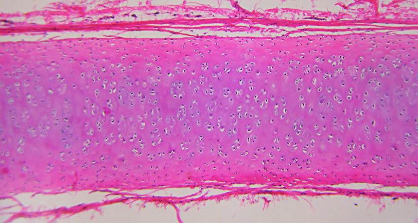
It has worked in goats. Now, in the first human trial, researchers at the University of Basel in Switzerland have grown cells called chondrocytes, taken from the noses of 10 patients with damaged knee joints, into cartilage grafts. These repair patches were then surgically implanted into the patients’ knee joints.

Two years after surgery, nine patients have seen improvements in knee function, quality of life and pain. (One patient dropped out of the trial because of additional athletic injuries.) MRI scans showed that the grafts looked like normal hyaline cartilage, the hard-to-replicate material that coats the tips of bones, the team reports in the Oct. 22 Lancet. Tests in more people are needed to determine whether the technique is ready for prime time.

Poor diet in pregnancy, poor heart health for infants

Mothers who don’t eat enough during pregnancy could give birth to babies with long-lasting heart problems. The results from a new study in primates add to accumulating evidence that a mother’s nutrition has more bearing on her child’s health than previously thought.

“We pass more biological milestones during development than we will ever pass again in our entire lives,” says Peter Nathanielsz, coauthor of the study published November 6 in the Journal of Physiology. And during those critical nine months, calorie intake at the extremes — too many or too few — appears to have a lifelong influence on newborn weight, future metabolism and chronic health problems (SN: 1/23/16, p. 22).
One landmark epidemiological investigation found that people born in the Dutch Hunger Winter during World War II suffered from an elevated risk of heart disease and other health concerns, with some risks even affecting two generations. But studies of human populations are complicated. It’s hard to account for the role of stress, behavior or environmental exposures. So Nathanielsz, of the University of Wyoming in Laramie, and colleagues from the University of Texas Health Science Center at San Antonio studied baboons, close genetic relatives to humans.

Sixteen pregnant baboons were fed their normal amount of chow, while 16 others received 30 percent less during pregnancy, a reduction researchers characterize as “moderate.” All other living conditions were the same. The researchers then compared offspring of the well-fed mothers with the offspring of undernourished mothers.

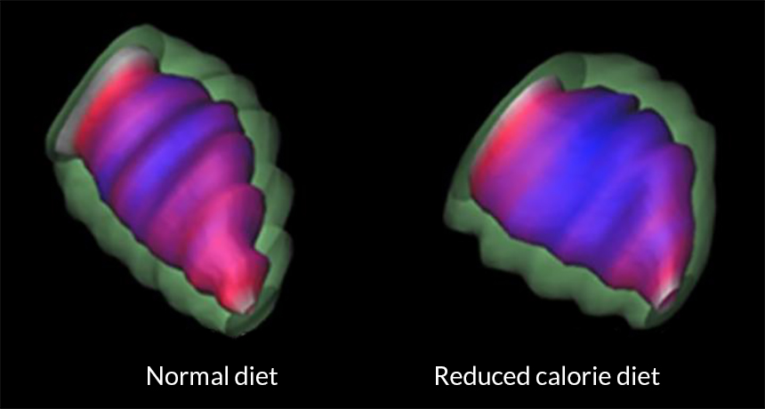
Infants of the underfed mothers were born small but nonetheless caught up in body weight to the offspring of the well-fed mothers by young adulthood. However, those whose mothers were underfed had more fibrous, abnormally shaped heart muscle, the researchers report. A normal heart is roughly an upside-down pyramid, but underfed offspring had more rounded and less muscular hearts. Evidence showed that these less-muscled hearts were not as efficient at pumping blood, with an average output about 20 percent lower.

Offspring undernourished in the womb also had hearts that appeared to age faster. By age five, the human equivalent of almost 25, many of their heart functions more closely resembled those of hearts of primates about three times as old.

Such experiments can show cause and effect — something that human studies can’t do, says Susan Ozanne, a developmental endocrinologist at the University of Cambridge. As a result, they provide strong evidence about the effects of maternal nutrition. “What this shows us is that certainly maternal diet has an effect on a child’s cardiac health long-term,” she says. Studies in rodents have produced similar findings, but “when you validate those in multiple species, it shows you you’re looking at a fundamentally conserved mechanism.”
The next step, she says, is to learn whether diet and exercise after birth can make up for poor nutrition during development. Doctors also don’t know whether there is a window of time during childhood for intervention, or a longer period to counteract any effects, she says.

Much attention on maternal nutrition has focused on the obesity epidemic, Nathanielsz says, but undernutrition remains a public health challenge throughout the world, even in developed countries. The U.S. Department of Agriculture estimates that approximately 13 percent of American households in 2015 reported food insecurity, or uncertainty about having enough money for food. “The number of people with food insecurity is very high,” Nathanielsz says. “It would be sad if we discounted this problem.”

Readers respond to the SN 10, and awards for Science News

Science’s human side
In “The SN 10: Scientists to Watch” (SN: 10/1/16, p. 16), Science News recognized 10 up-and-coming scientists across a range of scientific fields who will be answering big questions in the decades to come.

Barry Maletzky thought that highlighting 10 young scientists may have been unfair and detrimental to other researchers. “By drawing attention to just 10, I wonder if you are thereby discouraging others who may not make the headlines but whose basic research may lead the way toward important discoveries in the future,” Maletzky said. He also pointed out that scientific progress is made “through the tedious and often under-publicized work of a number of investigators working barely noticed through the years.”
It’s true that in science journalism, as with all journalism, what you choose to cover can matter as much as how you choose to cover it, says Elizabeth Quill, Science News’ enterprise editor, who led the SN 10 project. Science News editors and writers take this responsibility seriously, which is why Science News avoided terms like “top,” “best” and other superlatives. Whenever possible, the names of mentors and collaborators were also included in the stories. “We recognize the dangers in calling out specific individuals, but we believe the rewards outweigh the risks,” Quill says. “The majority of Science News focuses on the data and the process. But here we see a different side. We are showing science as a human endeavor. We hope this list inspires all young scientists to follow their passion and their curiosity. But we also hope to do what we do best — inform our readers about new and interesting science.”

Kenneth Abate was disappointed by “The SN 10: Scientists to Watch” profiles and questioned how Science News chose the researchers. “The issue is probably a nice piece of advertising for the individuals to further their careers,” Abate said. However, it “is of little value to the scientific community nor does it contribute to the knowledge bank of those scientists or would-be scientist readers.”
Choosing is never easy, but thankfully Science News staff didn’t do it alone. Every featured scientist was nominated by a Nobel laureate or recently elected member of the National Academy of Sciences on the basis of the scientist’s contributions to the field and promise for future contributions. As they do with any news or feature story, Science News editors and writers selected the final list of scientists by looking at who was doing novel, interesting and important work. As readers point out, the list could have easily been much, much longer.

Cool it
The next big thing in high-tech clothing may be a plastic material similar to kitchen cling wrap that vents body heat, Meghan Rosen reported in “New fabric could make cool clothes” (SN: 10/1/16, p. 9).

Online reader Karl Chwe pointed out one potential drawback to the new material: “It is just a thin plastic film with tiny holes, and the holes aren’t big enough to allow water vapor to escape easily, so it doesn’t allow evaporative cooling,” he wrote. Chwe suggested that it might be better to wear fewer clothes.

Actually, the nanopores are permeable to water vapor, the authors reported. “In this regard, the new fabric is comparable to cotton,” Rosen says. “Without the nanopores, the fabric would be a literal sweat suit; it’s completely nonpermeable.” But even with pores, the fabric doesn’t quite feel like normal clothes just yet. Weaving the fibers into a textile could help. Still, when it comes to evaporative cooling, Rosen says, wearing fewer clothes might be the simplest solution of all.

Correction
“A gut check gets personal” (SN: 10/1/16, p. 19) profiles Lawrence David, a computational biologist who studies the human gut microbiome at Duke University. David’s wife is a psychiatrist, not a psychologist as was incorrectly stated in the article.

Blue leaves help begonias harvest energy in low light

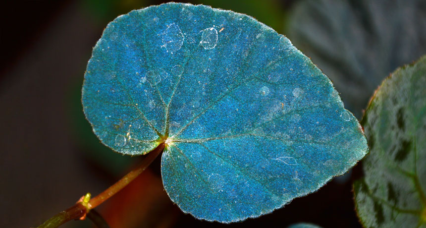
Iridescent blue leaves on some begonias aren’t just for show — they help the plants harvest energy in low light.

The begonias’ chloroplasts, which use photosynthesis to convert light into fuel, have a repeating structure that allows the plants to efficiently soak up light. This comes in handy for a plant that lives on the shady forest floor. The structure acts as a “photonic crystal” that preferentially reflects blue wavelengths of light and helps the plant better absorb reds and greens for energy production, scientists report October 24 in Nature Plants.
Colors in plants and animals typically come from pigments, chemicals that absorb certain wavelengths, or colors, of light. In rare cases, plants and animals derive their hues from microstructures. In begonias, such tiny, regular architectures can be found within certain chloroplasts, known as iridoplasts. As light bounces off these structures within an iridoplast, the reflected waves interfere at certain wavelengths (SN: 6/7/08, p. 26), creating a blue, iridescent shimmer.
Those structured chloroplasts also offer a survival benefit, the new research shows: They help the plants collect light. In a hybrid of two species — Begonia grandis and Begonia pavonina — the structures enhance the absorption of green and red wavelengths by concentrating these rays on light-absorbing compartments within the iridoplasts. Importantly, the structures slow the light. The “group velocity,” or the speed of a packet of light waves, is decreased due to interference between incoming and reflected light. The slowdown gives the plant more time to absorb precious sunbeams.

“These iridoplasts can basically photosynthesize at low-light levels where normal chloroplasts just simply could not photosynthesize,” says study coauthor Heather Whitney, a plant biologist at the University of Bristol in England. Iridoplasts, however, can’t hold their own in bright light. So begonias also have standard chloroplasts, which provide energy in plentiful sunshine. Iridoplasts act like “a backup generator” in dim conditions, Whitney says.

Other plants have structured chloroplasts, too, so begonias might not be alone in their feats of light manipulation. “Plants can’t really run away from their problems,” Whitney says. Instead, they have to be crafty enough to survive where they stand.

Having an extra chromosome has a surprising effect on cancer

SAN FRANCISCO — Having an extra chromosome may suppress cancer, as long as things don’t get stressful, a new study suggests. The finding may help scientists unravel a paradox: Cells with extra chromosomes grow slower than cells with the usual two copies of each chromosome, but cancer cells, which grow quickly, often have additional chromosomes. Researchers have thought that perhaps extra chromosomes and cancer-causing mutations team up to produce tumors.

Jason Sheltzer, a cell biologist at Cold Spring Harbor Laboratory in New York, and colleagues examined the effect of having an extra chromosome in mouse cells that also have cancer-promoting mutations. Cells with an extra copy of a chromosome — known as trisomic cells — grew slower in lab dishes and formed smaller tumors in mice than cells with cancer mutations but no extra chromosomes. Even when trisomic cells carry cancer-associated genes on the extra chromosome, the cells make less than usual of the cancer-driving proteins produced from those genes, Sheltzer reported December 5 at the annual meeting of the American Society for Cell Biology.

Extra chromosomes aren’t entirely off the hook for promoting cancer, though. After cells carrying extra chromosomes were grown with a low dose of chemotherapy drugs, they grew faster than cells that don’t have extra chromosomes, Sheltzer discovered. That could be because cells remaining after chemotherapy have developed additional abnormalities that might make the cancer more aggressive, he said.

Year in review: Ozone hole officially on the mend

In a rare bright spot for global environmental news, atmospheric scientists reported in 2016 that the ozone hole that forms annually over Antarctica is beginning to heal. Their data nail the case that the Montreal Protocol, the international treaty drawn up in 1987 to limit the use of ozone-destroying chemicals, is working.

The Antarctic ozone hole forms every Southern Hemisphere spring, when chemical reactions involving chlorine and bromine break apart the oxygen atoms that make up ozone molecules. Less protective ozone means that more ultraviolet radiation reaches Earth, where it can damage DNA and lead to higher rates of skin cancer, among other threats.
The Montreal Protocol cut back drastically on the manufacture of ozone-destroying compounds such as chlorofluorocarbons, or CFCs, which had been used in air conditioners, refrigerators and other products. It went into force in 1989 and phased out CFCs by 2010.

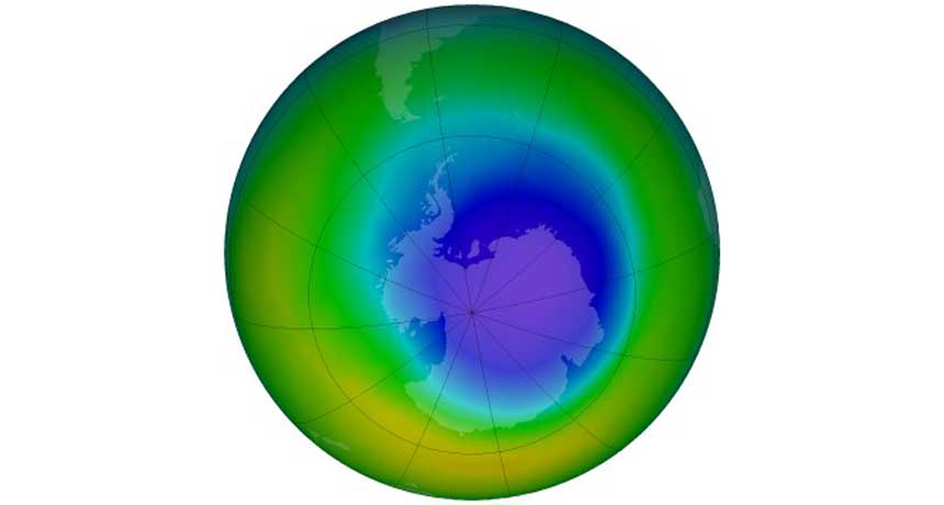
Earlier studies had hinted that the ozone hole was on the mend. The new work, reported in Science in June, is the most definitive yet (SN: 7/23/16, p. 6). A team led by Susan Solomon, an atmospheric chemist at MIT, looked not only at the month of October, when Antarctic ozone loss typically peaks, but also at September, when the hole is growing. The healing trend was most obvious in September. Satellite measurements showed that from 2000 to 2015, the average extent of the September ozone hole shrank by about 4.5 million square kilometers, to approximately 18 million square kilometers. Soundings taken by weather balloons over Antarctica confirmed the findings.
CFC concentrations peaked above Antarctica in the late 1990s and early 2000s and have been dropping ever since, says Birgit Hassler, an atmospheric chemist at Bodeker Scientific in Alexandra, New Zealand. Each passing year allows scientists to gather more convincing data. The new study, Hassler says, “makes the whole development of the Antarctic ozone hole healing very transparent and understandable.”
It is a fitting capstone to Solomon’s career. In the 1980s she led a team that proposed that chlorine compounds were to blame for Antarctic ozone loss. She then traveled to the frozen continent to conduct pioneering experiments that measured the accumulating chemicals there. “It’s very humbling now to be 30 years later and be able to say we have a clear fingerprint that the ozone hole is starting to get better,” she says.

Solomon says that public engagement was key to solving the ozone problem, with people coming together to identify an issue that threatened society and develop new technologies to fix it. In that respect, the most successful environmental treaty in history holds lessons for dealing with a much bigger threat, she says — climate change.

To fix the ozone layer, industry stopped using CFCs and similar compounds and replaced them with hydrofluorocarbons. Those chemicals, however, turned out to be powerful greenhouse gases that accelerated global warming. In October, the nations that ratified the Montreal Protocol agreed to expand it to cover hydrofluorocarbons as well (SN: 11/26/16, p. 13).

For some salamanders, finding a mate is a marathon

When looking for love, some small-mouthed salamanders can really go the distance.

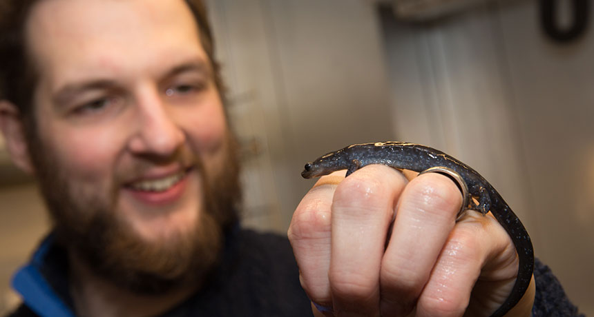
These intrepid amphibians (Ambystoma texanum) will risk death and dehydration to travel almost nine kilometers on average and as far as 14 to find a mate, researchers report December 20 in Functional Ecology. But all-female populations of a closely related group of salamanders that reproduce by cloning can’t go nearly as far.

Scientists tested the amphibians’ endurance on tiny treadmills. Then the team analyzed genetic differences between salamanders in patches of Ohio wetlands to see how far the amphibians might roam in the wild. Unisexual salamanders could only go a quarter of the treadmill distance that the small-mouthed salamanders could. And in the wild, they only dispersed about half as far from the pools where they were born.
By making the treacherous trek to a different pool to mate, A. texanum salamanders can mix up their genes and keep healthy variation in each population. Unisexual salamanders may have less stamina because they don’t mate in the usual way. Instead of searching for the perfect partner, they steal sperm from nearby male salamanders of different species. The sperm kick-start egg production but rarely actually fertilize eggs; only occasionally does a male’s DNA sneak into a female’s offspring.

Ditching the guys can be efficient — every member of an all-female population can give birth, and that means more babies. But it seems that going it alone has drawbacks, too: These salamanders’ poorer endurance could be a disadvantage if environmental changes forced them to colonize new territory.

New form of hydrogen created

Scientists have produced a new form of hydrogen in the lab — negatively charged hydrogen clusters.

Each cluster consists of hydrogen molecules arranged around a negatively charged hydrogen ion — a single hydrogen atom with an extra electron — at temperatures near absolute zero, the researchers report in the Dec. 30 Physical Review Letters. Similar, positively charged ion clusters have previously been found, but this is the first time scientists have seen negative hydrogen cluster ions beyond the simplest possible pairing of one molecule and one ion.
Physicist Michael Renzler of the University of Innsbruck in Austria and colleagues infused tiny droplets of liquid helium with hydrogen gas. Then, the scientists bombarded the droplets with a beam of electrons, which converted some hydrogen molecules into negatively charged hydrogen ions. Neighboring hydrogen molecules (two bonded hydrogen atoms) clustered around the ion, in groups of a few molecules to over 60.

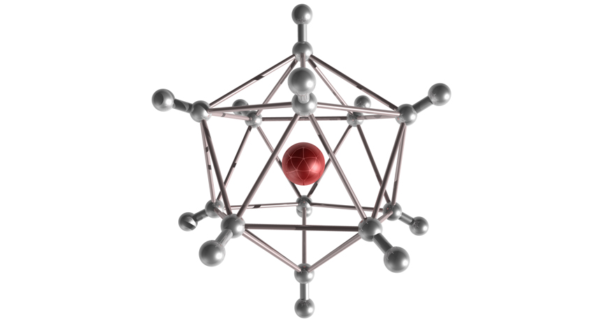
The scientists also determined the geometric structures of the clusters. Hydrogen molecules organized into shells that surrounded the central ion. Clusters were most stable, and most common, when molecules filled shells to their capacity. In the first shell, for example, the cluster formed an icosahedron — a 3-D shape with 12 vertices — when 12 molecules filled this shell.

In space, hydrogen cluster ions might form naturally in cold, dense clouds of hydrogen or in atmospheres of gas giant planets.